CAZypedia celebrates the life of Senior Curator Emeritus Harry Gilbert, a true giant in the field, who passed away in September 2025.
CAZypedia needs your help!
We have many unassigned pages in need of Authors and Responsible Curators. See a page that's out-of-date and just needs a touch-up? - You are also welcome to become a CAZypedian. Here's how.
Scientists at all career stages, including students, are welcome to contribute.
Learn more about CAZypedia's misson here and in this article. Totally new to the CAZy classification? Read this first.
Difference between revisions of "Auxiliary Activity Family 3"
| Line 38: | Line 38: | ||
Cellobiose dehydrogenases (CDHs) are extracellular flavocytochromes that were first described in 1974 <cite>Westermark1974</cite>. CDHs are exclusively found in wood-degrading and phytopathogenic fungi belonging to the phyla Basidiomycota (Class-I CDHs) and Ascomycota (Class-II and -III CDHs) <cite>Kracher2016b</cite>. They oxidize a wide variety of lignocellulose-derived saccharides to their corresponding sugar lactones. CDHs show a high preference for soluble, β-(1,4)-interlinked saccharides and scarcely oxidize monosaccharides. | Cellobiose dehydrogenases (CDHs) are extracellular flavocytochromes that were first described in 1974 <cite>Westermark1974</cite>. CDHs are exclusively found in wood-degrading and phytopathogenic fungi belonging to the phyla Basidiomycota (Class-I CDHs) and Ascomycota (Class-II and -III CDHs) <cite>Kracher2016b</cite>. They oxidize a wide variety of lignocellulose-derived saccharides to their corresponding sugar lactones. CDHs show a high preference for soluble, β-(1,4)-interlinked saccharides and scarcely oxidize monosaccharides. | ||
| − | A common feature of all CDHs is their complex bipartite structure, which comprises a C-terminal cytochrome-binding domain (CYT) and a larger, catalytic flavodehydrogenase (DH) domain encoded within a single polypeptide chain <cite>Hallberg2002</cite> (Figure 2A). Both domains are connected by a flexible linker which typically comprises 15 – 30 amino acids. An important in vivo function of CDH is the reduction and activation of family [[AA9]] lytic polysaccharide monooxygenases via its heme ''b'' domain <cite>Phillips2011, Tan2015</cite>. Recently, the holoenzyme structures of ''Neurospora crassa'' CDH (pdb: [{{PDBlink}}4qi7 4QI7]) and ''Myriococcum thermophilum'' CDH (pdb: [{{PDBlink}}4qi6 4QI6]) were reported. Two structures of ''N. crassa'' CDH showed an “open-state” conformation in which DH and CYT were spatially separated, whereas a structure of ''M. thermophilum'' CDH showed a “closed-state” conformation in which the propionate arm of the cytochrome domain interacted with the catalytic centre in DH <cite> Tan2015</cite>. Analysis by small angle scattering also suggested a number of possible intermediate conformers that exist in solution <cite>Tan2015, Bodenheimer2018</cite>. While the closed-state allows interdomain electron transfer from DH to CYT, reduction of electron acceptors (e.g. [[AA9]] enzymes) might occur in the open-state, in which the heme cofactor is fully accessible. | + | A common feature of all CDHs is their complex bipartite structure, which comprises a C-terminal cytochrome-binding domain (CYT) and a larger, catalytic flavodehydrogenase (DH) domain encoded within a single polypeptide chain <cite>Hallberg2002</cite> (Figure 2A). Both domains are connected by a flexible linker which typically comprises 15 – 30 amino acids. An important in vivo function of CDH is the reduction and activation of family [[AA9]] lytic polysaccharide monooxygenases via its heme ''b'' domain <cite>Phillips2011, Tan2015 Courtade2016</cite>. Recently, the holoenzyme structures of ''Neurospora crassa'' CDH (pdb: [{{PDBlink}}4qi7 4QI7]) and ''Myriococcum thermophilum'' CDH (pdb: [{{PDBlink}}4qi6 4QI6]) were reported. Two structures of ''N. crassa'' CDH showed an “open-state” conformation in which DH and CYT were spatially separated, whereas a structure of ''M. thermophilum'' CDH showed a “closed-state” conformation in which the propionate arm of the cytochrome domain interacted with the catalytic centre in DH <cite> Tan2015</cite>. Analysis by small angle scattering also suggested a number of possible intermediate conformers that exist in solution <cite>Tan2015, Bodenheimer2018</cite>. While the closed-state allows interdomain electron transfer from DH to CYT, reduction of electron acceptors (e.g. [[AA9]] enzymes) might occur in the open-state, in which the heme cofactor is fully accessible. |
| Line 82: | Line 82: | ||
#Phillips2011 pmid=22004347 | #Phillips2011 pmid=22004347 | ||
#Tan2015 pmid=26151670 | #Tan2015 pmid=26151670 | ||
| + | #Courtade2016 pmid=27152023 | ||
#Bodenheimer2018 pmid=29374564 | #Bodenheimer2018 pmid=29374564 | ||
#Fernandez2009 pmid=19923715 | #Fernandez2009 pmid=19923715 | ||
Revision as of 09:09, 2 May 2018
This page is currently under construction. This means that the Responsible Curator has deemed that the page's content is not quite up to CAZypedia's standards for full public consumption. All information should be considered to be under revision and may be subject to major changes.
- Author: ^^^Roland Ludwig^^^ and ^^^Daniel Kracher^^^
- Responsible Curator: ^^^Roland Ludwig^^^
| Auxiliary Activity Family AA3 | |
| Clan | AA3 |
| Mechanism | FAD-dependent substrate oxidation |
| Active site residues | known |
| CAZy DB link | |
| https://www.cazy.org/AA3.html | |
General properties and substrate specificities

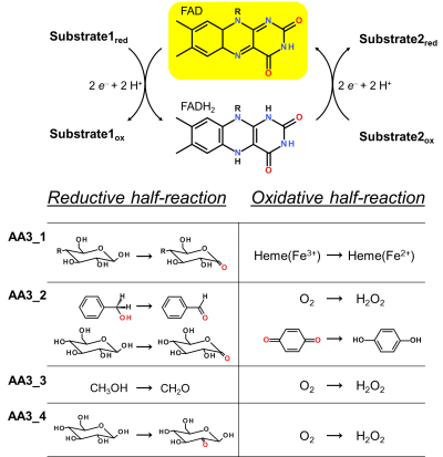
Enzymes of the CAZy family AA3 are widespread and catalyse the oxidation of alcohols or carbohydrates with the concomitant formation of hydrogen peroxide or hydroquinones [1]. AA3 enzymes are most abundant in wood-degrading fungi where they typically display a high multigenicity [2]. The main function of fungal AA3 enzymes is the stimulation of lignocellulose degradation in cooperation with other AA-enzymes such as peroxidases (AA2) [3] or lytic polysaccharide monooxygenases (AA9) [4, 5]. In yeast, AA3 enzymes are involved in the catabolism of alcohols [6], and some AA3 genes identified in insects are thought to be relevant for immunity and development [7, 8]. Recently, a bacterial AA3 enzyme with unknown biological function was isolated and characterized [9]. The functionally diverse enzymes of family AA3 all belong to the structurally related glucose-methanol-choline (GMC) family of oxidoreductases and require a flavin-adenine dinucleotide (FAD) cofactor for catalytic activity. Based on their sequences members of the AA3 family were divided into four subfamilies in the CAZy database (Figure 1). Family AA3_1 contains the flavodehydrogenase domain of cellobiose dehydrogenase (EC 1.1.99.18), family AA3_2 includes aryl alcohol oxidase (EC 1.1.3.7), glucose oxidase (EC 1.1.3.4), glucose dehydrogenase (EC 1.1.5.9) and pyranose dehydrogenase (EC 1.1.99.29), family AA3_3 consists of alcohol (methanol) oxidases (EC 1.1.3.13) and family AA3_4 comprises pyranose oxidoreductases (EC 1.1.3.10).
Subfamily AA3_1: cellobiose dehydrogenase
Cellobiose dehydrogenases (CDHs) are extracellular flavocytochromes that were first described in 1974 [10]. CDHs are exclusively found in wood-degrading and phytopathogenic fungi belonging to the phyla Basidiomycota (Class-I CDHs) and Ascomycota (Class-II and -III CDHs) [11]. They oxidize a wide variety of lignocellulose-derived saccharides to their corresponding sugar lactones. CDHs show a high preference for soluble, β-(1,4)-interlinked saccharides and scarcely oxidize monosaccharides. A common feature of all CDHs is their complex bipartite structure, which comprises a C-terminal cytochrome-binding domain (CYT) and a larger, catalytic flavodehydrogenase (DH) domain encoded within a single polypeptide chain [12] (Figure 2A). Both domains are connected by a flexible linker which typically comprises 15 – 30 amino acids. An important in vivo function of CDH is the reduction and activation of family AA9 lytic polysaccharide monooxygenases via its heme b domain [13, 14, 15]. Recently, the holoenzyme structures of Neurospora crassa CDH (pdb: 4QI7) and Myriococcum thermophilum CDH (pdb: 4QI6) were reported. Two structures of N. crassa CDH showed an “open-state” conformation in which DH and CYT were spatially separated, whereas a structure of M. thermophilum CDH showed a “closed-state” conformation in which the propionate arm of the cytochrome domain interacted with the catalytic centre in DH [14]. Analysis by small angle scattering also suggested a number of possible intermediate conformers that exist in solution [14, 16]. While the closed-state allows interdomain electron transfer from DH to CYT, reduction of electron acceptors (e.g. AA9 enzymes) might occur in the open-state, in which the heme cofactor is fully accessible.
Subfamily AA3_2
Aryl Alcohol Oxidase/Dehydrogenase

Aryl alcohol oxidoreductases (AAO) are secreted by basidiomycete fungi and proposed to play a crucial role in lignin degradation. The X-ray crystal structure of the most widely studied AAO from Pleurotus eryngii (pdb: 3FIM) showed a non-covalently bound FAD cofactor in the active-site [17] (Figure 2C). Access to the active site is restricted by three aromatic residues, which interact with both the alcohol substrate and oxygen. The substrate scope of AAO includes secreted benzyl alcohols from the secondary metabolism (e.g. veratryl alcohol), or related alcohols that accumulate during lignin degradation [3]. During the reductive half-reaction, the primary alcohol of the substrate is two-electron oxidized to form the corresponding aldehyde. In addition, aromatic aldehydes can undergo further oxidation yielding the corresponding acids. The concomitantly formed hydrogen peroxide is considered essential for the activity of lignin degrading peroxidases. Recently, aryl-alcohol quinone oxidoreductases (AAQO) with a high sequence identity to Pleurotus eryngii AAO were identified in the genome of the basidiomycete Pycnoporus cinnabarinus [18]. While these enzymes showed a substrate spectrum similar to known AAOs, oxygen reduction was insignificant or very low relative to other characterised AAOs. However, reoxidation of FADH2 was very efficient with benzoquinone or phenoxy-radicals, which are formed by fungal laccases, suggesting a different functional role of AAQOs.
Glucose oxidase and glucose dehydrogenase
Glucose oxidases (GOX) (Figure 2D) and glucose dehydrogenases (GDH) catalyse the regioselective oxidation of β-D-glucose to D-glucono-1,5-lactone. Both GOX and GDH are structurally related, including a set of conserved active site residues for the binding of glucose. Therefore, a discrimination of these genes based on their sequences is difficult [2]. Glucose oxidases typically occur as homodimers whereas GDHs occur as monomers or dimers. The first crystal structure of GOX from Aspergillus niger was resolved in 1993 (pdb: 1GAL [19]). A highly conserved arrangement of active-site residues, which form hydrogen bonds to all five hydroxyl groups of β-D-glucose, is responsible for the particularly high specificity towards Dglucose. The first crystal structure of GDH from Aspergillus flavus was reported in 2013, showing a slightly less conserved active site and fewer interactions with glucose (pdb: 4YNT [20]). This may also explain the promiscuous reaction of GDH with β-D-xylose, which is not observed for GOX. Both enzymes show different cosubstrate specificities. Glucose oxidases reduce atmospheric oxygen to hydrogen peroxide in their oxidative half-reaction. Proposed native functions of the enzyme are closely related to its peroxide-producing abilities and include the preservation of honey, microbial defence as well as the support of H2O2-dependent ligninases in wood degrading fungi [21]. GDHs interact very slowly with oxygen and prefer electron acceptors like quinones or phenoxy-radicals. The detailed physiological role of GDH remains elusive, but has been previously linked to the neutralization of plant laccase activity by fungi during plant infection [22].
Pyranose dehydrogenase
Pyranose dehydrogenase (PDH) is a secretory protein found in a limited number of litter degrading fungi belonging to the families Agaricaceae and Lycoperdaceae [23]. While the overall sequential and structural similarities to other GMC-oxidoreductases are high, the enzyme differs in terms of its catalytic properties and contains a covalently linked FAD, which is not observed in other AA3_2 enzymes. PDHs are characterised by a broad substrate spectrum, which includes a number of monosaccharides (L-arabinose, D-glucose, and D-galactose, D-xylose) as well as some oligo- or polysaccharides. PDHs catalyse the C1, C2, or C3 oxidation of the sugar substrate, or introduce di-oxidations at C2/C3 or C3/C4, resulting in the formation of aldonolactones (C1 oxidation) or (di)ketosugars. The first crystal structure of the enzyme was resolved in 2013 (pdb: 4H7U) and showed an open active site conformation, which in part may also explain its high substrate promiscuity [24]. The electron acceptor specificity is similar to those of other sugar dehydrogenases and includes quinones and phenoxy radicals.
Subfamily AA3_3: alcohol oxidase
Alcohol- or methanol oxidases (AOX) (Figure 2D) are intra- or extracellular enzymes found in yeasts and fungi. In methylotrophic yeasts, such as Pichia pastoris, AOX is a catabolic enzyme essential for the utilization of methanol. AOX catalyses the selective oxidation of primary alcohols at CH-OH, yielding the corresponding carbonyl compounds [25]. The enzyme accepts both saturated and unsaturated aliphatic alcohols with a chain length from C1 to C8, but shows a strong discrimination against secondary alcohols. To date, two structures of AOX from P. pastoris were resolved in 2016; One based on X-ray diffraction (pdb: 5HSA [26]), the other on cyro-electron microscopy (pdb: 5I68 [27]). AOX are homo-octameric enzymes located in the peroxisomal matrix [25]. Each subunit contains a covalently bound FAD-molecule modified with an arabinyl chain. In P. pastoris, the degree of the FAD arabinylation depends on the alcohol concentration in the medium and is thought to modify the reactivity with methanol. Considerably less is known about fungal AOX, which have been found in wood-degrading or phytopathogenic fungi from the phylum Basidiomycota. Similarly to AOX from yeast, they showed an octameric architecture and displayed the highest catalytic efficiencies for methanol. The fungal AOX from Gloeophyllum trabeum was identified as extracellular protein despite the lack of a dedicated secretion signal peptide [28]. The native function of fungal AOX could therefore be related to its peroxide production, which, similar to other AA3_2 enzymes, can potentially stimulate fungal attack on lignocellulose by supplying H2O2 to peroxidases. AOX secreted by the phytopathogenic basidiomycete Moniliophthora perniciosa, the causative agent of Witches’ broom disease in the cocoa tree, is thought to play a role in the utilization of methanol derived from pectin demethylation [29].
Subfamily AA3_4: pyranose oxidase
Pyranose oxidases (POX) (Figure 2B) are widespread in lignocellulose degrading fungi and catalyse the C2-oxidation of monosaccharides. POX is the most distantly related member of the AA3 family and does not show the strict conservation of structural motifs found within the AA3 family. Unlike other AA3 enzymes, POXs are associated with membrane-bound vesicles and other membrane structures in the periplasmic space of the fungal hyphae. The first crystal structure of POX was reported in 2004 (Trametes multicolor, pdb: 1TT0 [30]). The enzyme is a homo-tetramer with each of the subunits carrying a covalently bound FAD molecule. A distinct “head domain” on each of the subdomains is thought to be involved in oligomerization or in interactions with cell wall-polysaccharides [30]. Access to the active site of the enzyme is modulated by a flexible active-site loop, which hinders entrance of oligosaccharides. Substrate channels lead from the polypeptide surface to an internal large cavity formed by the four subunits. The preferred substrate of POX is either α- or β-D-glucose, but also D-galactose, D-xylose, or D-glucono-1,5-lactone are converted [31]. Substrates are oxidized at the C2 position to yield 2-ketoaldoses as products. In the oxidative half-reaction, the FADH2 is regenerated by reduction of O2 to H2O2. The reduction of oxygen by POX proceeds through a C4a-hydroperoxyflavin intermediate, which is characteristic for FAD-dependent monooxygenases but is untypical for sugar oxidases [32]. Apart from oxygen, POX can also utilize alternative electron acceptors including a number of (substituted) quinones and (complexed) metal ions. Of note, catalytic efficiencies for some of these electron acceptors are higher than for oxygen, suggesting that these acceptors might be biologically more relevant substrates than oxygen. Recently, putative POX sequences were identified in Actinobacteria with overall sequence identities of 39–24% to fungal POX [9]. A functional POX from Arthrobacter siccitolerans could be recombinantly expressed and characterized [9]. POX activity was also confirmed in the supernatant of the bacterium Pantoea ananatis when cultivated on rice straw [33].
References
Error fetching PMID 22249717:
Error fetching PMID 27127235:
Error fetching PMID 27312718:
Error fetching PMID 23525937:
Error fetching PMID 17498303:
Error fetching PMID 23022604:
Error fetching PMID 12493734:
Error fetching PMID 22004347:
Error fetching PMID 26151670:
Error fetching PMID 27152023:
Error fetching PMID 29374564:
Error fetching PMID 19923715:
Error fetching PMID 26873317:
Error fetching PMID 8421298:
Error fetching PMID 26311535:
Error fetching PMID 18330562:
Error fetching PMID 21903757:
Error fetching PMID 11511865:
Error fetching PMID 23326459:
Error fetching PMID 16169288:
Error fetching PMID 26905908:
Error fetching PMID 27458710:
Error fetching PMID 17660304:
Error fetching PMID 23022488:
Error fetching PMID 15288786:
Error fetching PMID 11472941:
Error fetching PMID 18652479:
Error fetching PMID 27761153:
- Levasseur A, Drula E, Lombard V, Coutinho PM, and Henrissat B. (2013). Expansion of the enzymatic repertoire of the CAZy database to integrate auxiliary redox enzymes. Biotechnol Biofuels. 2013;6(1):41. DOI:10.1186/1754-6834-6-41 |
- Error fetching PMID 29411063:
- Error fetching PMID 22249717:
- Error fetching PMID 27127235:
- Error fetching PMID 27312718:
- Error fetching PMID 23525937:
- Error fetching PMID 17498303:
- Error fetching PMID 23022604:
-
Mendes S, Banha C, Madeira J, Santos D, Miranda V, Manzanera M, Ventura MR, van Berkel WJH, Martins LO. Characterization of a bacterial pyranose 2-oxidase from Arthrobacter siccitolerans. 2016 J Mol Catal B Enzym 133, S34–S43
-
Westermark U, Eriksson KE. Cellobiose:Quinone Oxidoreductase, a New Wood-degrading Enzyme from White-rot Fungi. 1974 Acta Chem Scand 1974 28b:209–214.
-
Kracher D, Ludwig R. Cellobiose dehydrogenase: An essential enzyme for lignocellulose degradation in nature - A review. 2016 Bodenkultur 67:145–163.
- Error fetching PMID 12493734:
- Error fetching PMID 22004347:
- Error fetching PMID 26151670:
- Error fetching PMID 27152023:
- Error fetching PMID 29374564:
- Error fetching PMID 19923715:
- Error fetching PMID 26873317:
- Error fetching PMID 8421298:
- Error fetching PMID 26311535:
- Error fetching PMID 18330562:
- Error fetching PMID 21903757:
- Error fetching PMID 11511865:
- Error fetching PMID 23326459:
- Error fetching PMID 16169288:
- Error fetching PMID 26905908:
- Error fetching PMID 27458710:
- Error fetching PMID 17660304:
- Error fetching PMID 23022488:
- Error fetching PMID 15288786:
- Error fetching PMID 11472941:
- Error fetching PMID 18652479:
- Error fetching PMID 27761153: