CAZypedia celebrates the life of Senior Curator Emeritus Harry Gilbert, a true giant in the field, who passed away in September 2025.
CAZypedia needs your help!
We have many unassigned pages in need of Authors and Responsible Curators. See a page that's out-of-date and just needs a touch-up? - You are also welcome to become a CAZypedian. Here's how.
Scientists at all career stages, including students, are welcome to contribute.
Learn more about CAZypedia's misson here and in this article. Totally new to the CAZy classification? Read this first.
Difference between revisions of "Glycoside Hydrolase Family 128"
Harry Brumer (talk | contribs) m (minor grammatical improvements) |
|||
| Line 29: | Line 29: | ||
== Substrate specificities == | == Substrate specificities == | ||
| − | The first GH128 enzyme, GLU1, was cloned from ''Lentinula edodes'' fruiting bodies (shiitake mushroom) <cite>Sakamoto2011</cite>. GLU1 cleaves β-1,3 linkages in various β-glucans such as lentinan | + | The first GH128 enzyme, GLU1, was cloned from ''Lentinula edodes'' fruiting bodies (shiitake mushroom) <cite>Sakamoto2011</cite>. GLU1 cleaves β-1,3 linkages in various β-glucans such as endogenous 'L. edodes'' lentinan, laminarin from ''Laminaria digitata'', pachyman from ''Poria cocos'', and curdlan from ''Alcaligenes faecalis'', but does not degrade β-1,3-linkages within β-1,3-1,4-glucans such as barley glucan, indicating the enzyme is categorized into EC [{{EClink}}3.2.1.39 3.2.1.39] <cite>Sakamoto2011</cite>. Further work with several GH128 members corroborated that this family is specific for β-1,3-glucans <cite>Santos2020</cite>. In addition, it was demonstrated that bacterial members, such as those from ''Amycolatopsis mediterranei'' (subgroup I) and ''Pseudomonas viridiflava'' (subgroup II), are endo-β-1,3-glucanases that degrade these carbohydrates at higher rates <cite>Santos2020</cite>. Fungal GH128 members are more diverse in terms of activity: endo-β-1,3-glucanases, represented by the GLU1 from ''L. edodes'' (subgroup IV) <cite>Sakamoto2011 Santos2020</cite>; exo-β-1,3-glucanases that release trisaccharides (''Aureobsidium namibiae'') (subgroup VI) <cite>Santos2020</cite> and monosaccharides (''Cryptococcus neoformans'') (subgroup V) from the reducing ends; and exo-β-1,3-glucanases that release trisaccharides from the non-reducing ends of triple-helical β-1,3-glucans, represented by the enzyme from ''Blastomyces gilchristii'' (subgroup III) <cite>Santos2020</cite>. Some fungal members from this family, such as those from ''Trichoderma gamsii'' and ''C. neoformans'', are devoid of catalytic activity but conserve the capacity to bind short β-1,3-glucooligosaccharides (subgroup VII) <cite>Santos2020</cite>. |
| + | |||
== Kinetics and Mechanism == | == Kinetics and Mechanism == | ||
| − | As | + | As indicated by the first study of a GH128 enzyme <cite>Sakamoto2011</cite>, this family is part of [[Clan]] GH-A, thus suggesting that its members operate by a classical Koshland [[retaining]] mechanism. This prediction was confirmed through <sup>1</sup>H-nuclear magnetic resonance spectroscopy of enzymatic products <cite>Santos2020</cite>. |
== Catalytic Residues == | == Catalytic Residues == | ||
| − | From the sequence alignment of GH128 members, two glutamic acids, E103 and E195 in ''L. edodes'' GLU1, were predicted to be the catalytic residues <cite>Sakamoto2011</cite>. They were further confirmed to be the acid/base and the nucleophile, respectively, by site-directed mutagenesis of the bacterial GH128 member from ''A. mediterranei'' <cite>Santos2020</cite>. These residues are located at the C-terminal ends of the strands β7 and β4 <cite>Santos2020</cite>, as observed for other clan GH-A families. | + | From the sequence alignment of GH128 members, two glutamic acids, E103 and E195 in ''L. edodes'' GLU1, were predicted to be the catalytic residues <cite>Sakamoto2011</cite>. They were further confirmed to be the [[general acid/base]] and the [[catalytic nucleophile]], respectively, by site-directed mutagenesis of the bacterial GH128 member from ''A. mediterranei'' <cite>Santos2020</cite>. These residues are located at the C-terminal ends of the strands β7 and β4 <cite>Santos2020</cite>, as observed for other [[clan]] GH-A families. |
== Three-dimensional structures == | == Three-dimensional structures == | ||
Revision as of 07:19, 17 August 2020
This page is currently under construction. This means that the Responsible Curator has deemed that the page's content is not quite up to CAZypedia's standards for full public consumption. All information should be considered to be under revision and may be subject to major changes.
- Author: ^^^Yuichi Sakamoto^^^ and ^^^Camilla Santos^^^
- Responsible Curator: ^^^Mario Murakami^^^
| Glycoside Hydrolase Family GH128 | |
| Clan | GH-A |
| Mechanism | retaining |
| Active site residues | known |
| CAZy DB link | |
| https://www.cazy.org/GH128.html | |
Substrate specificities
The first GH128 enzyme, GLU1, was cloned from Lentinula edodes fruiting bodies (shiitake mushroom) [1]. GLU1 cleaves β-1,3 linkages in various β-glucans such as endogenous 'L. edodes lentinan, laminarin from Laminaria digitata, pachyman from Poria cocos, and curdlan from Alcaligenes faecalis, but does not degrade β-1,3-linkages within β-1,3-1,4-glucans such as barley glucan, indicating the enzyme is categorized into EC 3.2.1.39 [1]. Further work with several GH128 members corroborated that this family is specific for β-1,3-glucans [2]. In addition, it was demonstrated that bacterial members, such as those from Amycolatopsis mediterranei (subgroup I) and Pseudomonas viridiflava (subgroup II), are endo-β-1,3-glucanases that degrade these carbohydrates at higher rates [2]. Fungal GH128 members are more diverse in terms of activity: endo-β-1,3-glucanases, represented by the GLU1 from L. edodes (subgroup IV) [1, 2]; exo-β-1,3-glucanases that release trisaccharides (Aureobsidium namibiae) (subgroup VI) [2] and monosaccharides (Cryptococcus neoformans) (subgroup V) from the reducing ends; and exo-β-1,3-glucanases that release trisaccharides from the non-reducing ends of triple-helical β-1,3-glucans, represented by the enzyme from Blastomyces gilchristii (subgroup III) [2]. Some fungal members from this family, such as those from Trichoderma gamsii and C. neoformans, are devoid of catalytic activity but conserve the capacity to bind short β-1,3-glucooligosaccharides (subgroup VII) [2].
Kinetics and Mechanism
As indicated by the first study of a GH128 enzyme [1], this family is part of Clan GH-A, thus suggesting that its members operate by a classical Koshland retaining mechanism. This prediction was confirmed through 1H-nuclear magnetic resonance spectroscopy of enzymatic products [2].
Catalytic Residues
From the sequence alignment of GH128 members, two glutamic acids, E103 and E195 in L. edodes GLU1, were predicted to be the catalytic residues [1]. They were further confirmed to be the general acid/base and the catalytic nucleophile, respectively, by site-directed mutagenesis of the bacterial GH128 member from A. mediterranei [2]. These residues are located at the C-terminal ends of the strands β7 and β4 [2], as observed for other clan GH-A families.
Three-dimensional structures
A three-dimensional homology model of L. edodes GLU1 indicated similarity with several (β/α)8-barrel (TIM-barrel) structures, including a GH39 β-xylosidase and a GH5 β-mannanase [1]. The fold resembling an (β/α)8-barrel was further confirmed with the crystal structure determination of 9 members of the family [2]. However, in all structures, the helix α2 and the strand β3 are strictly absent [2]. Moreover, some enzymes such as the endo-β-1,3-glucanase from ‘‘L. edodes’’ (GLU1) and the exo-β-1,3-glucanase from C. neoformans, also lack the helices α1 and α3, respectively [2].
Two distinct modes of substrate binding were observed in the GH128 family [2]. The most widespread mode, named as hydrophobic knuckle, involves a tryptophan residue that interacts with four glucoside moieties from –5 to –2 and is fully complementary to the typically curved conformation of β-1,3-glucan chains. The other mode, only observed in fungal members belonging to subgroups IV and VI, requires substrate conformational changes to allow the binding to the catalytic interface. In these fungal subgroups, the hydrophobic knuckle is absent and two aromatic residues, positioned at the -5 and -4 subsites, create a linearized cleft, which requires a 180° torsion in the glycosidic bond between the glycosyl moieties –2 and –3 in the β-1,3-glucan chain for binding. This mode of substrate recognition is called as “flattening” mechanism due to the unusual conformational, but also stereochemically favorable, adopted by the substrate. It is notable that such mode of substrate binding was not yet observed in other CAZy families active on β-1,3-glucans.
Clustering of GH128

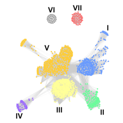
The glycoside hydrolase family 128 was created based on the study of Yuichi Sakamoto and colleagues [1]. Years later, the group headed by Mario Murakami carried out a task force to explore the functional and structural diversity of this family [2]. For this purpose, they employed phylogenetic and SSN analyses to segregate the family into putative isofunctional subgroups. The SSN analysis resulted in two well discretized clusters (subgroups VI and VII) and a third cluster that was further subdivided into five subgroups (I to V) based on SSN alignment scores and evolutionary closeness (Fig. 1). At least one member of each subgroup was biochemically and structurally characterized: AmGH128_I, PvGH128_II, ScGH128_II, BgGH128_III, LeGH128_IV, CnGH128_V, AnGH128_VI, TgGH128_VII and CnGH128_VII. Subgroups I and II were found to be predominantly present in bacteria, and the subgroups III to VII are mostly found in fungi. Bacterial enzymes are faster, present the hydrophobic knuckle and attack the β-1,3-glucan in an endo mode of action, which is compatible with their biological function: nutrition and competition. Fungal β-1,3-glucanases are known to act on remodeling of their own cell walls. Therefore, these enzymes are slower, more diverse in terms of substrate recognition modes (flattening mechanism – subgroups IV and VI; hydrophobic knuckle – subgroups III, V and VII) and mode of action (exo-enzymes – subgroups III, V and VI; endo-enzymes – subgroup IV; oligosaccharide binding proteins – subgroup VII). This was the first time that a glycoside hydrolase family was rationally studied based on SSN analysis. A recent study led by Prof. Harry Brumer applied a similar strategy to classify the polyspecific GH16 family into isofunctional subgroups using the available functional and structural data in the literature [3], highlighting this approach as a promising strategy to systematically assess the functional and structural diversity of CAZyme families. It is noteworthy to point out that Brumer´s group made available an intuitive and robust program to perform SSN analyses, named as SSNpipe that is freely available from GitHub (https://github.com/ahvdk/SSNpipe).
Family Firsts
- First stereochemistry determination
- predicted to be retaining by membership in Clan GH-A [1] and further validated by 1H-NMR of products of the A. mediterranei endo-β-1,3-glucanase (AmGH128_I) [2].
- First catalytic nucleophile identification
- predicted by sequence alignment [1] and confirmed by site-directed mutagenesis of A. mediterranei endo-β-1,3-glucanase (AmGH128_I) [2].
- First general acid/base residue identification
- predicted by sequence alignment [1] and confirmed by site-directed mutagenesis of A. mediterranei endo-β-1,3-glucanase (AmGH128_I) [2].
- First 3-D structure
- predicted by modelling of L. edodes GLU1 [1] and experimentally determined for several GH128 members including endo-β-1,3-glucanases from A. mediterranei (AmGH128_I), P. viridiflava (PvGH128_II), Sorangium cellulosum (ScGH128_II) and L. edodes (LeGH128_IV); exo-β-1,3-glucanases from B. gilchristii (BgGH128_III), C. neoformans (CnGH128_V) and A. namibiae (AnGH128_VI); and β-1,3-glucooligosaccharide binding proteins from T. gamsii (TgGH128_VII) and C. neoformans (CnGH128_VII) [2].
References
- Sakamoto Y, Nakade K, and Konno N. (2011). Endo-β-1,3-glucanase GLU1, from the fruiting body of Lentinula edodes, belongs to a new glycoside hydrolase family. Appl Environ Microbiol. 2011;77(23):8350-4. DOI:10.1128/AEM.05581-11 |
- Santos CR, Costa PACR, Vieira PS, Gonzalez SET, Correa TLR, Lima EA, Mandelli F, Pirolla RAS, Domingues MN, Cabral L, Martins MP, Cordeiro RL, Junior AT, Souza BP, Prates ÉT, Gozzo FC, Persinoti GF, Skaf MS, and Murakami MT. (2020). Structural insights into β-1,3-glucan cleavage by a glycoside hydrolase family. Nat Chem Biol. 2020;16(8):920-929. DOI:10.1038/s41589-020-0554-5 |
- Viborg AH, Terrapon N, Lombard V, Michel G, Czjzek M, Henrissat B, and Brumer H. (2019). A subfamily roadmap of the evolutionarily diverse glycoside hydrolase family 16 (GH16). J Biol Chem. 2019;294(44):15973-15986. DOI:10.1074/jbc.RA119.010619 |