CAZypedia celebrates the life of Senior Curator Emeritus Harry Gilbert, a true giant in the field, who passed away in September 2025.
CAZypedia needs your help!
We have many unassigned pages in need of Authors and Responsible Curators. See a page that's out-of-date and just needs a touch-up? - You are also welcome to become a CAZypedian. Here's how.
Scientists at all career stages, including students, are welcome to contribute.
Learn more about CAZypedia's misson here and in this article. Totally new to the CAZy classification? Read this first.
Difference between revisions of "Glycoside Hydrolase Family 189"
Harry Brumer (talk | contribs) |
Harry Brumer (talk | contribs) |
||
| Line 33: | Line 33: | ||
The reaction products of TiCGS<sub>Cy</sub> were obtained using LβG as the substrate. The β-glucosidase-resistant compounds in the products were purified by size-exclusion chromatography. <sup>1</sup>H-NMR analysis of these purified polysaccharides suggested that they are CβGs. In the chemical shift of <sup>1</sup>H-NMR, no chemical shift derived from an anomeric proton in an α-anomer moiety was detected. This observation clearly demonstrates that TiCGS<sub>Cy</sub> has a retaining mechanism <cite>Tanaka2024</cite>. | The reaction products of TiCGS<sub>Cy</sub> were obtained using LβG as the substrate. The β-glucosidase-resistant compounds in the products were purified by size-exclusion chromatography. <sup>1</sup>H-NMR analysis of these purified polysaccharides suggested that they are CβGs. In the chemical shift of <sup>1</sup>H-NMR, no chemical shift derived from an anomeric proton in an α-anomer moiety was detected. This observation clearly demonstrates that TiCGS<sub>Cy</sub> has a retaining mechanism <cite>Tanaka2024</cite>. | ||
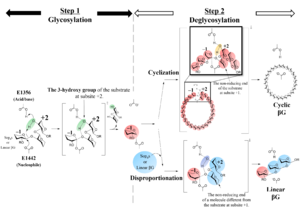
| − | Structural analysis (see [[#Three- | + | Structural analysis (see [[#Three-dimensional Structure]] below) and mutational analysis suggest that E1442 of TiCGS<sub>Cy</sub> acts on an anomeric carbon of a glucose moiety at subsite −1 as a nucleophile and E1356 of TiCGS<sub>Cy</sub> acts on a scissile bond of a substrate via 3-hydroxy group of a glucose moiety at subsite +2 as an acid/base <cite>Tanaka2024</cite>. The reaction mechanism of TiCGS<sub>Cy</sub> is noncanonical in that a substrate hydroxy group participates in the catalytic process (Fig.1)<cite>Tanaka2024</cite>.[[Image:Fig1.png.png|thumb|widthpx|'''Fig.1''' '''Catalytic mechanism of TiCGS<sub>Cy</sub>.''' ]] |
== Catalytic Residues == | == Catalytic Residues == | ||
Revision as of 17:27, 24 February 2024
This page has been approved by the Responsible Curator as essentially complete. CAZypedia is a living document, so further improvement of this page is still possible. If you would like to suggest an addition or correction, please contact the page's Responsible Curator directly by e-mail.
- Authors: Tomoko Masaike, Masahiro Nakajima, and Nobukiyo Tanaka
- Responsible Curator: Masahiro Nakajima
| Glycoside Hydrolase Family GH189 | |
| Clan | GH-x |
| Mechanism | Retaining |
| Active site residues | Known |
| CAZy DB link | |
| https://www.cazy.org/GH189.html | |
Substrate specificities
The cyclization domain alone of cyclic β-1,2-glucan synthase from Thermoanaerobacter italicus (TiCGSCy) was identified, characterized and structurally analyzed as reported in 2024 [1]. This enzyme established the novel glycoside hydrolase family (GH) 189. This enzyme specifically catalyzes transglycosylation reactions on linear β-1,2-glucans (LβGs) and β-1,2-glucooligosaccharides (Sopns, where 'n' represents the degree of polymerization (DP)) with DP 6 or more [1]. In the deglycosylation step, intermolecular transglycosylation results in release of disproportionated linear products, while intramolecular transglycosylation results in cyclization of the substrates to release cyclic β-1,2-glucans (CβGs) [1].
Kinetics and Mechanism
The reaction products of TiCGSCy were obtained using LβG as the substrate. The β-glucosidase-resistant compounds in the products were purified by size-exclusion chromatography. 1H-NMR analysis of these purified polysaccharides suggested that they are CβGs. In the chemical shift of 1H-NMR, no chemical shift derived from an anomeric proton in an α-anomer moiety was detected. This observation clearly demonstrates that TiCGSCy has a retaining mechanism [1].
Structural analysis (see #Three-dimensional Structure below) and mutational analysis suggest that E1442 of TiCGSCy acts on an anomeric carbon of a glucose moiety at subsite −1 as a nucleophile and E1356 of TiCGSCy acts on a scissile bond of a substrate via 3-hydroxy group of a glucose moiety at subsite +2 as an acid/base [1]. The reaction mechanism of TiCGSCy is noncanonical in that a substrate hydroxy group participates in the catalytic process (Fig.1)[1].
Catalytic Residues
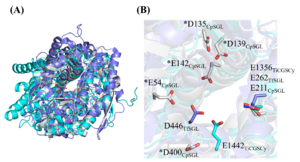
The acid/base residue and the nucleophile residue of TiCGSCy are E1356 and E1442, respectively. In addition, these residues are well superimposed with the general acid and the nucleophilic water in the GH162 fungal β-1,2-glucanase from Talaromyces funiculosus (TfSGL), respectively [1, 2]. TfSGL has an inverting mechanism particularly catalyzing a unique reaction via the 3-hydroxy group of the glucose molecule at subsite +2 [2].
As of Jan. 2024, the detailed reaction mechanisms of GH144 enzymes remain unclear [3]. However, SGLs belonging to both GH144 and GH162 have an inverting mechanism unlike GH189 [1, 2, 3]. A structural comparison revealed that the positions of the acid/base residue in GH189, the candidate catalytic residues in GH144 and the general acid residue in GH162 are well superimposed. On the other hand, the positions of the other catalytic residues (or candidate catalytic residues) in GH189, GH144 and GH162 are completely different (Fig.2).
Three-dimensional structure
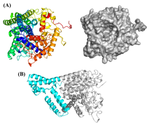
The apo-structure of the recombinant TiCGSCy was determined at 3.8 Å by X-ray crystal structure analysis (PDB: 8WY1) [1]. The overall structure comprises a single (α/α)6-barrel domain with several inserted α-helices and TiCGSCy has a large active-site pocket (Fig.3) [1].
Interestingly, although GH189 (= TiCGSCy), GH144 and GH162 enzymes are quite different in their amino acid sequences, their overall structures and the positions of the substrate in their catalytic pockets are similar [1].
GH144 and GH162 were officially classified as part of clan GH-S in this paper [1]. Although GH189 enzymes share a similar (α/α)6 fold structure with GH144 and GH162 enzymes, GH189 enzymes have a retaining mechanism unlike GH144 and GH162 enzymes with an inverting mechanism. Due to the fundamental difference in reaction mechanisms, GH189 is not included in clan GH-S; instead, GH189 could be regarded as a member of a potential new GH clan [1].
Family Firsts
- First stereochemistry determination
- 1H-NMR analysis of β-glucosidase-resistant compounds, which produced by TiCGSCy using LβG as a substrate, as described above [1].
- First catalytic nucleophile identification
- The structural and mutational analysis of TiCGSCy [1].
- First general acid/base residue identification
- The structural and mutational analysis of TiCGSCy [1].
- First three-dimensional structure
- The apo-structure of the recombinant TiCGSCy was determined by X-ray crystal structure analysis [1].
References
Error fetching PMID 30926603:
Error fetching PMID 28270506:
- Error fetching PMID 38300345:
- Error fetching PMID 30926603:
- Error fetching PMID 28270506:


